mail_outline sales@mediastorehouse.com
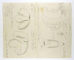
Diceros bicornis bicornis (Rhinoceros), organs, in or after 1778. Creator: Robert Jacob GordonDiceros bicornis bicornis (Rhinoceros), organs, in or after 1778
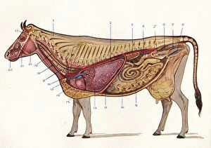
Median section of a cow, showing organs of circulation and respiration, etc, c1905 (c1910). From Live Stock in Health and Disease, edited by Professor J. Prince-Sheldon. [The Waverley Book Co
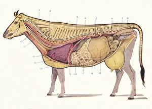
Median section of a cow, showing principal organs of digestion, etc, c1905 (c1910). From Live Stock in Health and Disease, edited by Professor J. Prince-Sheldon. [The Waverley Book Co
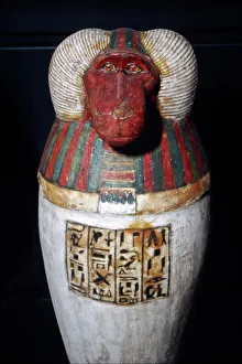
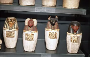
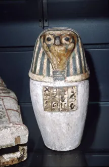
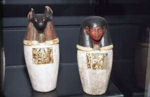
Thoth as Baboon, Canopic Jar, 22nd Dynasty, c1550BC-1069 BC. Containers used to store internal organs removed from the deceaseds body during mummification
Padiuf?s False Canopic Jars, 22nd Dynasty, c1550BC-1069 BC. Containers used to store internal organs removed from the deceaseds body during mummification
A vulture eating the innards of Titius, Cupid above two women to the left, set within and elaborate frame, from Loves, Rages and Jealousies of Juno, 1531-76
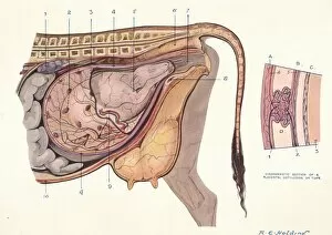
Section of the abdomen of a cow, showing foetus in normal position, c1905 (c1910)Section of the abdomen of a cow, showing foetus in normal position with diagrammatic section of a placental cotyledon or tuft, c1905 (c1910)
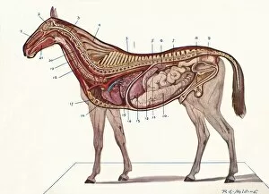
Vertical section of the body of a horse, c1907 (c1910). Artist: RE HoldingVertical section of the body of a horse, c1907 (c1910). From Live Stock in Health and Disease, edited by Professor J Prince-Sheldon. [The Waverley Book Co. Ltd, London, c1890]
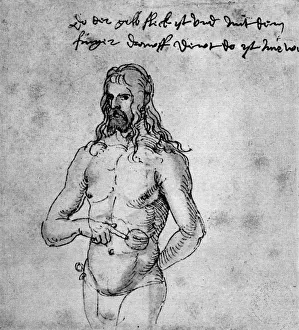
Self Portrait, about 1512-1513 or 1519, (1936). Artist: Albrecht DurerSelf Portrait, about 1512-1513 or 1519, (1936). Inscription: This is where the yellow spot is, an I am pointing to it with my finger: That is where it hurts
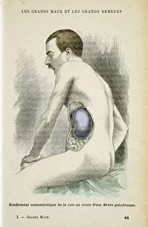
Typical enlarged spleen of a Malaria patient, c1890. Malaria is caused a parasitic protozoa transmitted by the Anopheles mosquito
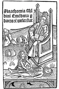
Anatomy demonstration, 1493. Title page of Anathomia by Mondino de Luzzi (Mundinus). Finished in 1316, Anathomia was first published in Padua in 1478
Osiris Canopic Jar, 22nd Dynasty, c1550BC-1069 BC. Containers used to store internal organs removed from the deceaseds body during mummification
Anubis Canopic Jars, 22nd Dynasty, c1550BC-1069 BC. Containers used to store internal organs removed from the deceaseds body during mummification
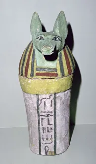
Jackal-headed wooden canopic jar for the storage of organs, Egyptian, 25th Dynasty, c700 BC. The head represents Duamutef, one of the four sons of Horus, and held the stomach
Canopic Jars from the Tomb of Tutankhamun. The heads represent the four protective goddesses Isis, Nephthys, Selket and Neith. Tutankhamun reigned between 1336 BC and 1327 BC
Foetus in uterus at time of quickening when first fetal movements are felt by the mother, c1795Foetus in the uterus at the time of quickening (16-18 weeks) when the first fetal movements are felt by the mother, c1795
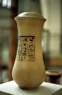
Canopic jar, vessel used for burial of embalmed viscera, Ancient Egyptian
Canopic Jars, Ancient Egyptian, 26th dynasty, 664-525 BC. These were used to contain the viscera of a dead person, usually for burial with their mummified body