mail_outline sales@mediastorehouse.com
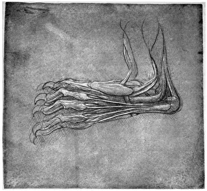
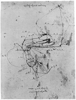
Muscles and sinews in a foot, possibly of a hare, late 15th or early 16th century (1954). Artist: Leonardo da VinciMuscles and sinews in a foot, possibly of a hare, late 15th or early 16th century (1954). Found in the collection of the Royal Library, Windsor Castle, Windsor, 12375
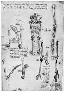
Study of human bones, late 15th or 16th century (1954). Artist: Leonardo da VinciStudy of human bones, late 15th or 16th century (1954). Found in the collection of the Royal Library, Windsor Castle, Windsor, 19004r. A print from Leonardo da Vinci by Ludwig H Heydenreich
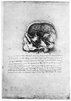
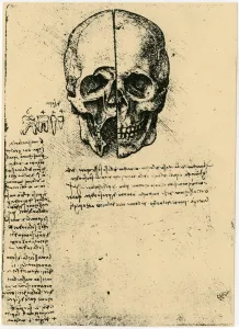
Study of a human skull, late 15th or early 16th century (1954). Artist: Leonardo da VinciStudy of a human skull, late 15th or early 16th century (1954). Found in the collection of the Royal Library, Windsor Castle, Windsor, 19058r. A print from Leonardo da Vinci by Ludwig H Heydenreich
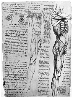
Muscle studies, late 15th or early 16th century (1954). Artist: Leonardo da VinciMuscle studies, late 15th or early 16th century (1954). Found in the collection of the Royal Library, Windsor Castle, Windsor, 19014v. A print from Leonardo da Vinci by Ludwig H Heydenreich
Study in proportion of a horses leg, late 15th or early 16th century (1954). Artist: Leonardo da VinciStudy in proportion of a horses leg, late 15th or early 16th century (1954). Found in the collection of the Royal Library, Windsor Castle, Windsor
Anatomy of a horse, 19th century. Artist: Archibald WebbAnatomy of a horse, 19th century
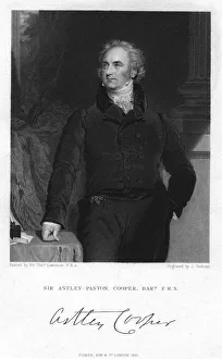
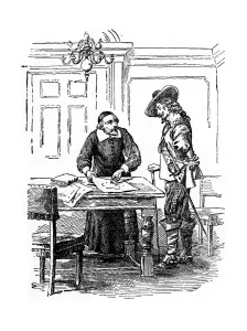
Sir Astley Paston Cooper, 1st Baronet, English surgeon and anatomist, 1831. Artist: J CochranSir Astley Paston Cooper, 1st Baronet, English surgeon and anatomist, 1831. Cooper (1768-1841) was Surgeon to Guys Hospital and a pupil of John Hunter
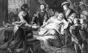
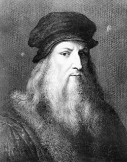
The death of Leonardo de Vinci, 1519. Artist: WalkerThe death of Leonardo de Vinci, 1519. One of the greatest figures of the Italian Renaissance, Leonardo (1452-1519) died at Clos Luce, Amboise, France, on 2nd May 1519
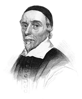
William Harvey, English physician, (c1850). Harvey (1578-1657) was a medical doctor who is credited with first correctly describing, in exact detail
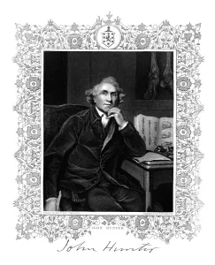
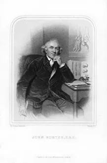
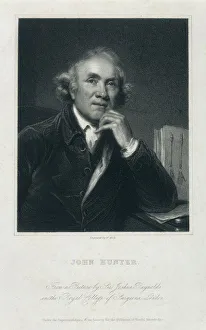
John Hunter, 18th century Scottish surgeon, (19th century). Artist: GH AdcockJohn Hunter, 18th century Scottish surgeon, (19th century). Hunter (1728-1793) delivered a unique series of lectures on the theory and practice of surgery, which attracted many famous students
William Harvey, 17th century English physician, (20th century). Harvey (1578-1657) pictured with King Charles I, his patron and friend
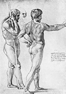
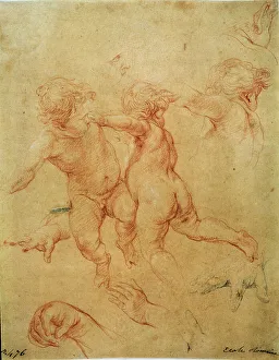
Two male nude studies, 1515, (1912). Artist: RaphaelTwo male nude studies, 1515, (1912). A drawing given by Raphael to Durer, with autograph of Raphael, and dated 1515. A print from The Connoisseur, (London, 1912)
John Hunter, Scottish surgeon, 1870. Artist: Francis HollJohn Hunter, Scottish surgeon, 1870. Hunter (1728-1793) gave a series of lectures on the theory and practice of surgery, which attracted many famous students, including Edward Jenner
Two flying putti, study, 1740s. Artist: Pompeo BatoniTwo flying putti, study, 1740s. Batoni, Pompeo Girolamo (1708-1787). Found in the collection of the State A. Pushkin Museum of Fine Arts, Moscow
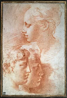
Study of the heads, c1527. Artist: ParmigianinoStudy of the heads, c1527. Parmigianino (1503-1540). Found in the collection of the State A. Pushkin Museum of Fine Arts, Moscow
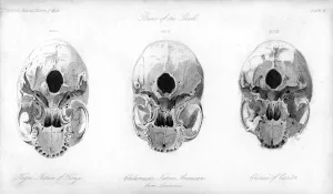
Base of the skull, 1848. Comparison of a Negro (native of Kongo), Chilamache native American (from Louisiana), and Chinese (from Canton)
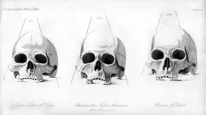
Three types of human skull, 1848. Comparison of a Negro (native of Kongo), Chilamache native American (from Louisiana), and Chinese (from Canton)
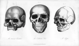
Ancient Cimbrian, Esthonian Finn, Lappe, 1848. An engraving from the Natural History of Man, by James Cowles Prichard, (Hippolyte Bailliere, London, third edition, 1848)
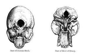
Human and orang-utan skulls, 1848. Illustrations of the base of the skull. An engraving from the Natural History of Man, by James Cowles Prichard, (Hippolyte Bailliere, London, third edition, 1848)
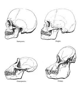
Lateral view of the skull or profile, 1848. Comparison of the skulls of European and African humans with those of the chimpanzee and orang-utan
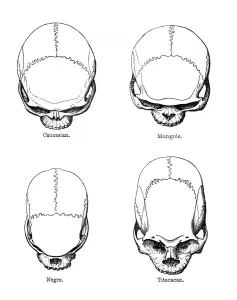
The vertical configuration of the skull, 1848. Comparison of Caucasian, Mongole, Negro and Titicacan skulls. During the 19th century
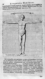
Proportions of man, 1675. Artist: Athanasius KircherProportions of man, 1675. A print from Arca Noe, Amsterdam, 1675. Found in the collection of Jean Claude Carriere
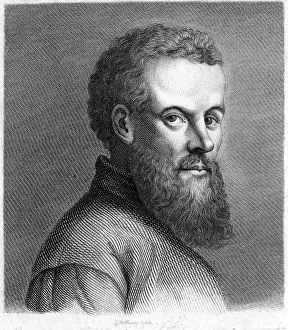
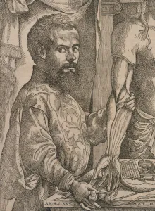
Andreas Vesalius, 16th century Flemish anatomist, c1789-c1798. Vesalius (1514-1564) great work on anatomy De Humani Corporis Fabrica (On the Structure of the Human Body) (1543) was a landmark
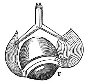
Descartes idea of the hydraulic action of the nerves, 1692. French philosopher Rene Descartes (1596-1650) believed nerves were hollow, provided with valves
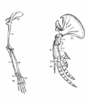
Homology (of commmon descent), c1920. A: Fore-limb of a monkey. B: Fore-limb of a whale. Although different at first sight, they have similar architecture
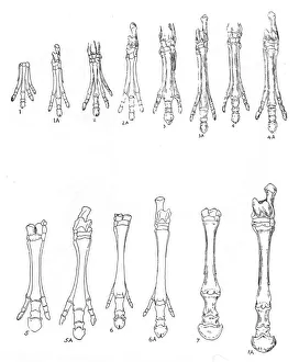
Evolution of the horse, c1920. Diagram of seven stages in the development of hind and forelimbs: 1, 1A Eohippus; 2, 2A Orohippus; 3, 3A Mesohippus; 4, 4A Hypohippus; 5, 5A Merychippus; 6
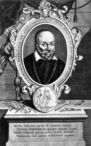
Girolamo Fabrici, Italian anatomist and surgeon, 17th century. Hieronymus Fabricius ab Aquapendente - Italian name Girolamo Fabrici - (1537-1619) was one of the founders of modern embryology
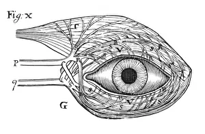
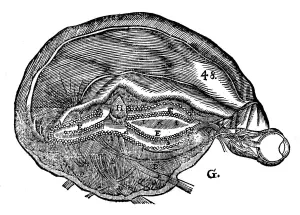
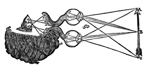
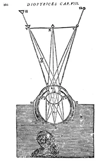
Rene Descartes diagram of the human brain and eye, 1692. From Opera Philosophica by Rene Descartes. (Frankfurt-am-Main, 1692). Originally published in his Tractatus de homine. (Paris, 1664)
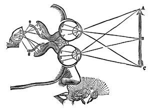
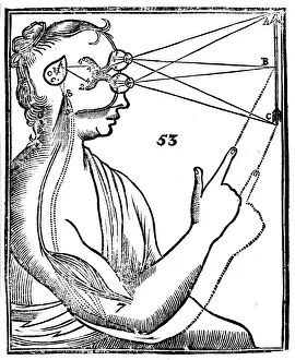
Rene Descartes illustration of the co-ordination of the senses, 1692. A visual stimulus travelling from the eye to the pineal gland, H, stops attention being given to an olfactory stimulus
Rene Descartes idea of vision, showing the function of the eye, optic nerve and brain, 1692. From Opera Philosophica by Rene Descartes. (Frankfurt-am-Main, 1692)
Descartes representation of the antagonistic eye muscles, 1692. When E is relaxed A is innervated. Rene Decartes (1596-1650)
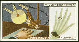
X-raying the hand, 1924. An X-ray tube and an X-ray photograph of a hand, with the bones and a wristwatch and ring clearly visible. Cigarette card
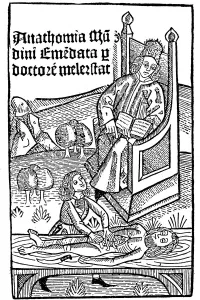
Anatomy demonstration, 1493. Title page of Anathomia by Mondino de Luzzi (Mundinus). Finished in 1316, Anathomia was first published in Padua in 1478
Descartes explanation of vision, 1692. Light rays being passed through the eye, being focused by the lens (I) and forming images T, S, R on the retina
Descartes idea of vision, 1692Descartes (1596-1650) idea of vision, [1692]. The passage of nervous impulses from the eye to the pineal gland and so to the muscles. From Rene Descartes Opera Philosophica (Tractatus de homine), 1692
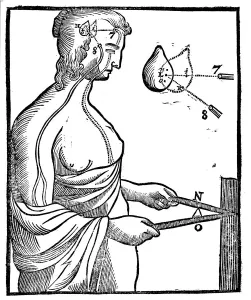
Descartes idea of how eye passes impulse to brain and so directs a voluntary movement, 1692. French philosopher and scientist Rene Descartes (1596-1650)
Involuntary movement, Descartes idea of how impulses from the limbs reach the brain, 1692. French philosopher and scientist Rene Descartes (1596-1650) believed all nerves to be hollow
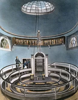
Theatre of Anatomy, Cambridge, 1815. From The History of the University of Cambridge, published by Ackermann. (London, 1815)
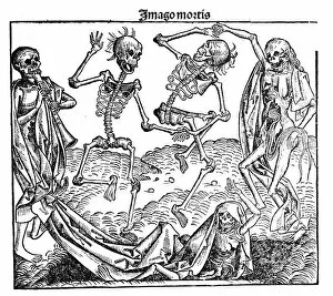
Death triumphant, 1493. From Liber chronicarum mundi (Nuremberg Chronicle), by Hartmann Schedel. The depiction of the skeletons is anatomically inaccurate, especially the bones of the pelvis
Circulation of the blood, 1628. English physician William Harvey (1578-1657) was the first to correctly describe the mechanism whereby blood is circulated in the body
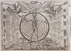
An astrological chart, 1646. Artist: Athanasius KircherAn astrological chart, 1646. A man with his internal organs revealed standing in front of a circular chart. One hand has been replaced by a sun
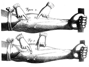
Andreas Vesalius dissecting the muscles of the forearm of a cadaver, 1543. Artist: Steven van CalcarAndreas Vesalius dissecting the muscles of the forearm of a cadaver, 1543. He exhibits a partly dissected arm of a taller man. Beside the arm, on the table, are instruments and a piece of text
John Hunter, FRS, (c1850-c1870?). Artist: William HollJohn Hunter, FRS, (c1850-c1870?). Portrait of the Scottish surgeon and anatomist. His unique series of lectures on the theory and practice of surgery attracted numerous students
Leonardo da Vinci, Italian artist, engineer, scientist and inventor, 1864Leonardo da Vinci, Italian artist, engineer, scientist and inventor whose drawings featured ideas such as a spinning wheel and a flying machine
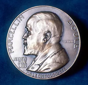
Pierre Marcellin Boule, French paleontologist, 20th century. In 1921 Boule (1861-1942) completed the first reconstruction of a Neanderthal skeleton. Obverse of commemorative medal
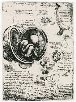
Anatomical sketch of a human foetus in the womb, c1510. Artist: Leonardo da VinciAnatomical sketch of a human foetus in the womb, c1510
Anatomical sketch of a human skull, c1472-1519. Artist: Leonardo da VinciAnatomical sketch of a human skull, c1472-1519
Anatomical sketch; two studies of a human skull, c1489. Artist: Leonardo da VinciAnatomical sketch; two studies of a human skull, c1489