mail_outline sales@mediastorehouse.com
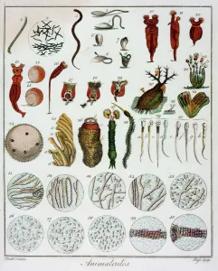
Animalcules observed by Anton van Leeuwenhoek, c1795. Hand-coloured engraving showing various items viewed through a microscope, including sperm
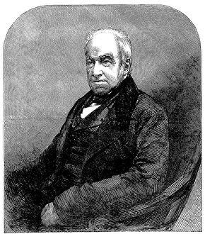
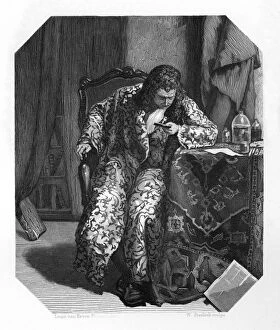
The Late Mr. Robert Brown, Keeper of Botany in the British Museum... 1858. Creator: UnknownThe Late Mr. Robert Brown, Keeper of Botany in the British Museum, from a photograph by Maull and Polyblank, 1858. Engraving from a photograph by Maull and Polyblank
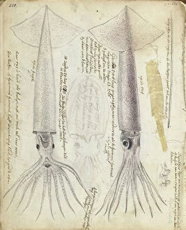
Cuttlefish and Gorita, 1785. Creator: Jan BrandesCuttlefish and Gorita, 1785. Sepia loligo'. Two colour drawings, and a sketch of an octopus-like creature. With inscriptions. Part of Jan Brandes sketchbook, dl. 1 (1808), p. 238
The Testimonial presented to Dr. Hassall, on Thursday, 1856. Creator: UnknownThe Testimonial presented to Dr. Hassall, on Thursday, 1856. Gift to British chemist and microscopist Arthur Hill Hassall
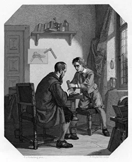
Hans Jansen and his son Sacharias, c1870. Artist: H SluyterHans Jansen and his son Sacharias, c1870. Some historians credit Sacharias Jansen, a Middelburg spectacle maker, with the invention of the telescope and the microscope
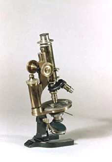
Microscope made by C Reichert, Vienna, 1895. Artist: C ReichertMicroscope made by C Reichert, Vienna, 1895. The microscope featured objectives of different powers which could be turned into viewing position as required
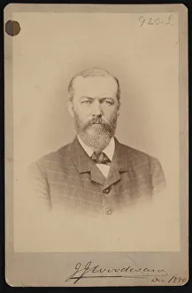
Portrait of Joseph Janvier Woodward (1833-1884), December 1880. Creator: UnknownPortrait of Joseph Janvier Woodward (1833-1884), December 1880
[Microscopic view of an insect], ca. 1853. Creator: Alois Auer[Microscopic view of an insect], ca. 1853
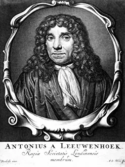
Antoni van Leeuwenhoek, 17th century Dutch scientist and microscopy pioneer, c1870. Artist: W SteelinkAntoni van Leeuwenhoek, 17th century Dutch scientist and microscopy pioneer, c1870. It was probably as a result of his use of lenses in examining cloth as a drapers apprentice that led to
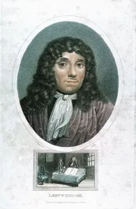
Antoni van Leeuwenhoek, Dutch pioneer of microscopy, (1813). Artist: J ChapmanAntoni van Leeuwenhoek, Dutch pioneer of microscopy, (1813). It was probably as a result of his use of lenses in examining cloth as a drapers apprentice that led to Leeuwenhoeks interest in lens
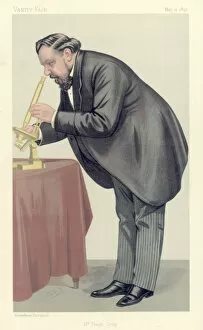
Mr Frank Crisp, 1890. Artist: SpyMr Frank Crisp, 1890. Crisp (c1853-1919), an English Limited Liability Lawyer one of whose personal interests was microscopy, acted as Secretary of the Royal Microscopical Society
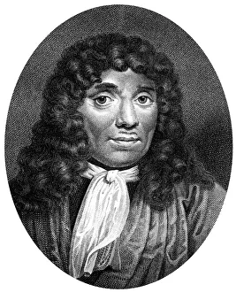
Frontispiece of Ontledigen en Ondekkigen... Brieven by Anton van Leeuwenhoek, 1686Frontispiece of Ontledigen en Ondekkigen...Brieven by Dutch microscopist Anton van Leeuwenhoek, 1686. Leeuwenhoek (1632-1723)
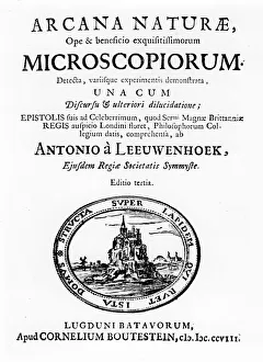
Title page of Microscopium by Dutch microscopist Anton van Leeuwenhoek, 1708. Leeuwenhoek (1632-1723) was one of the first to recognise cells in animals
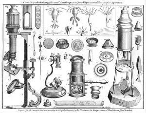
Microscopes and microscopical objects, 1750. I: Wilsons pocket microscope. II: Scroll microscope. III: Tripod microscope - improved form of Marshalls double microscope
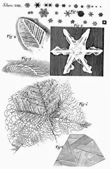
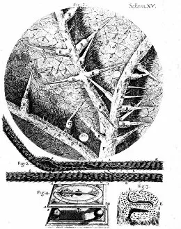
Frozen materials viewed by English microscopist Robert Hooke, 1665. Observations of several kinds of frozen figures showing frozen urine (1), snowflakes (2) and ice flakes (4, 5, 6)
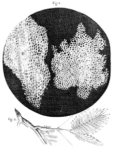
Hookes observations of the cellular structure of cork and a sprig of Sensitive Plant, 1665Hookes observations of the cellular structure of cork and a sprig of Sensible (Sensitive) Plant, 1665. Hooke was the first to use the word cell to describe the honeycomb nature of cork
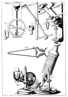
Hookes microscope with condenser for concentrating light, 1665. From left to right above are his barometer, refractometer for measuring refractive power of liquids, and lens-grinding machine
Illustrations from English microscopist Robert Hookes Micrographia, 1665. 1: underside of a stinging nettle leaf; 2: beard of wild oat used in Hookes hygrometer; 3: section of head of wild oat; 4
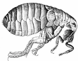
Flea, wingless bloodsucking parasitic insect, 1665. The human flea (Pulex irritans) can transmit plague. From Micrographia by Robert Hooke (1635-1703)
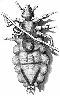
Louse clinging to a human hair, 1665Human Louse, a wingless parasitic insect, 1665. The human louse, a wingless parasitic insect, is now known to be a vector for epidemics of typhus. From Micrographia by Robert Hooke (1635-1703)
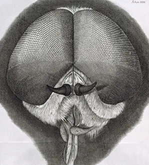
Grey drone-fly, observation XXXIX from Hookes Micrographia, 1664. Artist: Robert HookeGrey drone-fly, observation XXXIX from Hookes Micrographia, 1664. Micrographica contains prints of some of the specimens Hooke viewed under the compound microscope that he designed
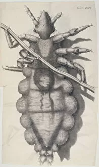
Louse clinging to a human hair in Hookes Micrographia, 1665. Robert Hooke was born on the Isle of Wight, and studied at Oxford University
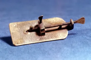
Microscope by Anton Van Leeuwenhoek, Dutch, c1670. Van Leeuwenhoek (1632-1723) was a Dutch scientist and microscopist who was the first to observe bacteria
Anton van Leeuwenhoek (1632-1723), Dutch microscopist, c1810. Artist: John ChapmanAnton van Leewenhoek (1632-1723) Dutch microscopist, c1810. It was probably as a result of his use of lenses in examining cloth as a drapers apprentice that led to Leeuwenhoeks interest in lens
Anton van Leeuwenhoek, Dutch pioneer of microscopy, 1723. Artist: Abraham de BloisAnton van Leeuwenhoek, Dutch pioneer of microscopy, c1660. It was probably as a result of his use of lenses in examining cloth as a drapers apprentice that led to Leeuwenhoeks interest in lens